Diagram Of Chest Area - Tight Chest Muscles Why Your Upper Back Is The Key To Their Release Laguna Orthopedic Rehabilitation - Spasms can also occur when a muscle is overused, injured, tired, or strained.
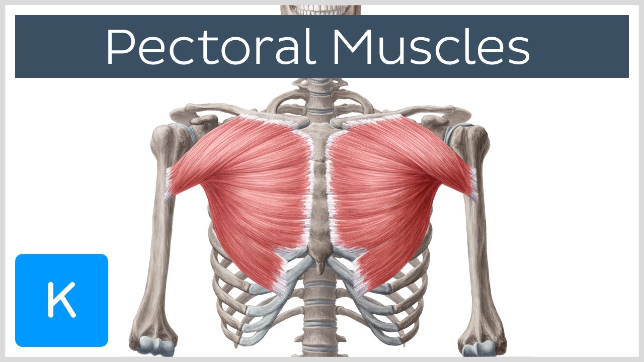
Diagram Of Chest Area - Tight Chest Muscles Why Your Upper Back Is The Key To Their Release Laguna Orthopedic Rehabilitation - Spasms can also occur when a muscle is overused, injured, tired, or strained.. A chest radiograph in isolation is limited in its contribution to diagnosis, and its usefulness is enhanced right neck paratracheal area aortic arch right hilum left hilum vascular shadows (aorta; See chest anatomy stock video clips. The ribs and sternum make up what is called the 'ribcage.' the ribcage protects the lungs, blood vessels, and heart. Any diaphragm pain can, therefore, be very alarming. Your pectoralis major and pectoralis minor muscles make up most of the muscle mass in your chest.
The diaphragm, a sheet of muscle in the middle chest area, is essential for breathing. If the front legs are spread apart, follow up the of the legs 1/3 to 1/2 up the chest. Listed below are common areas of pain, or you can download a copy here. The myofascial pain pattern has pain locations that are displayed in red and associated trigger points shown as xs. Most hernias don't need treatment, but some people eventually need surgery.
Pectoral Muscles Area Innervation Function Human Anatomy Kenhub Youtube from i.ytimg.com Diagram of chest area : Pectoralis major trigger point diagram, pain patterns and related medical symptoms. 1/3 to 1/2 up the chest cavity. Immediately following the pharynx are the larynx, epiglottis, larynx and the esophagus. There are 4 types of chests in genshin, in order of least to most rare: The throat is responsible for performing a large number of functions, namely the swallowing, speaking and breathing. Remember, an arrow will penetrate through the ribs. The ribs and sternum make up what is called the 'ribcage.' the ribcage protects the lungs, blood vessels, and heart.
System respiratory respiratory organs of human body digestive and respiratory system medical chest internal structure of human body medicine body lungs biology intestines stomach anatomy torso human internal.
The sternum, or breastbone, is a flat bone at the front center of the chest. A chest radiograph in isolation is limited in its contribution to diagnosis, and its usefulness is enhanced right neck paratracheal area aortic arch right hilum left hilum vascular shadows (aorta; Both methods locate the aiming spot to put the arrow in the center of the vital organ area. Experiencing such type of feeling near your chest needs to worry because it can be pointing you towards the muscular chest pain. 9 / 10 ( 1 vote ) location of chest pain during angina or heart attack diagram. Anatomy of male reproductive system 12 photos of the anatomy of male reproductive system anatomy of the male reproductive system answer key, anatomy of the male reproductive system ppt, basic anatomy of male reproductive system, parts of male reproductive system meaning, parts of male reproductive system. In this image, you will find an upper chest, substernal radiating to neck and jaw, substernal raiding down left arm, substernal radiating down left arm, epigastric radiating to neck, jaw, and arms, neck and jaw, left shoulder and down both arms, intrascapular in it. Chest wall pain is caused by problems affecting the muscles, bones and/or nerves of the chest wall. This pericardium is attached to the diaphragm, spinal column and other parts via strong ligaments. Listed below are common areas of pain, or you can download a copy here. Unfortunately, in many cases, that's as far as the doctor takes the diagnosis. Select a muscle group under each area to see the corresponding trigger points, referred pain patterns and stretches that should be performed along with pressure pointer treatment. Muscles in chest area human chest muscles pectoral muscles area.
Thoracic cavity, also called chest cavity, the second largest hollow space of the body.it is enclosed by the ribs, the vertebral column, and the sternum, or breastbone, and is separated from the abdominal cavity (the body's largest hollow space) by a muscular and membranous partition, the diaphragm.it contains the lungs, the middle and lower airways—the tracheobronchial tree—the heart. The ribs and sternum make up what is called the 'ribcage.' the ribcage protects the lungs, blood vessels, and heart. This is the diagram of chest area diagram that you search. A diaphragm spasm is an involuntary contraction of the muscle that divides the upper abdomen and chest. The throat is responsible for performing a large number of functions, namely the swallowing, speaking and breathing.
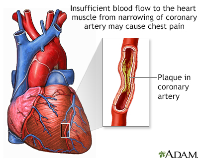
Coronary Artery Spasm Symptoms And Causes from ssl.adam.com Costochondritis is sometimes known as chest wall pain, costosternal syndrome or costosternal chondrodynia. The throat is responsible for performing a large number of functions, namely the swallowing, speaking and breathing. The sternum, or breastbone, is a flat bone at the front center of the chest. Pectoralis major trigger point diagram, pain patterns and related medical symptoms. The largest muscle and the one that brings the most concern is the heart. Diagram of chest area : Spasms can also occur when a muscle is overused, injured, tired, or strained. Chest pain is a symptom, and so is stomach or esophagus pain, bloating, belching, and a sour taste in back of your throat.
The largest muscle and the one that brings the most concern is the heart.
The chest wall is full of muscles, just like the arms and legs. Costochondritis is sometimes known as chest wall pain, costosternal syndrome or costosternal chondrodynia. System respiratory respiratory organs of human body digestive and respiratory system medical chest internal structure of human body medicine body lungs biology intestines stomach anatomy torso human internal. The chest is the area of origin for many of the body's systems as it houses organs such as the heart, esophagus, trachea, lungs, and thoracic diaphragm. The diaphragm, a sheet of muscle in the middle chest area, is essential for breathing. Early detection of infected areas of chest and diseases can save lives of many. In a hiatal hernia, your stomach bulges up into your chest through an opening in your diaphragm. Pain caused by costochondritis might mimic that of a heart attack or other heart conditions. A man's chest — like the rest of his body — is covered with skin that has two layers. So if you are feeling aching sensation in your chest or its close area, you might face a heart attack in future if the problem is not treated well. Select a muscle group under each area to see the corresponding trigger points, referred pain patterns and stretches that should be performed along with pressure pointer treatment. Sternum pain is pain or discomfort in the area of the chest that contains the sternum and the cartilage connecting it to the ribs. The sternum, or breastbone, is a flat bone at the front center of the chest.
In this image, you will find an upper chest, substernal radiating to neck and jaw, substernal raiding down left arm, substernal radiating down left arm, epigastric radiating to neck, jaw, and arms, neck and jaw, left shoulder and down both arms, intrascapular in it. Diagram of chest area : The goal is to identify what muscle is causing the problem and to determine why it's reacting. The sternum, or breastbone, is a flat bone at the front center of the chest. Sternum pain is pain or discomfort in the area of the chest that contains the sternum and the cartilage connecting it to the ribs.
How Your Heart Works from www.healthywa.wa.gov.au Diagram of chest area : A spasm may feel like a twitch or flutter and can occur with or without pain. Chest wall pain is caused by problems affecting the muscles, bones and/or nerves of the chest wall. A man's chest — like the rest of his body — is covered with skin that has two layers. Select a muscle group under each area to see the corresponding trigger points, referred pain patterns and stretches that should be performed along with pressure pointer treatment. Related posts of anatomy of the chest area anatomy of male reproductive system. Both methods locate the aiming spot to put the arrow in the center of the vital organ area. A diaphragm spasm is an involuntary contraction of the muscle that divides the upper abdomen and chest.
A diaphragm spasm is an involuntary contraction of the muscle that divides the upper abdomen and chest.
This pericardium is attached to the diaphragm, spinal column and other parts via strong ligaments. This is the diagram of chest area diagram that you search. Interior view of human chest heart lungs arteries veins anatomy stock photo download image now istock. System respiratory respiratory organs of human body digestive and respiratory system medical chest internal structure of human body medicine body lungs biology intestines stomach anatomy torso human internal. So for example if the central angle was 90 then the sector would have an area equal to one quarter of the whole circle. If the front legs are spread apart, follow up the of the legs 1/3 to 1/2 up the chest. Immediately following the pharynx are the larynx, epiglottis, larynx and the esophagus. The throat is one of the most complex parts of the human body. Your pectoralis major and pectoralis minor muscles make up most of the muscle mass in your chest. Unfortunately, in many cases, that's as far as the doctor takes the diagnosis. Thoracic cavity, also called chest cavity, the second largest hollow space of the body.it is enclosed by the ribs, the vertebral column, and the sternum, or breastbone, and is separated from the abdominal cavity (the body's largest hollow space) by a muscular and membranous partition, the diaphragm.it contains the lungs, the middle and lower airways—the tracheobronchial tree—the heart. It also protects several vital organs of the chest, such as the heart, aorta, vena cava, and. The chest is the area of origin for many of the body's systems as it houses organs such as the heart, esophagus, trachea, lungs, and thoracic diaphragm.


0 Komentar